COMPUTED TOMOGRAPHY (CT SCAN)
CT Scan
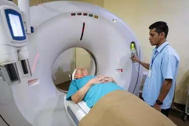
Computed Tomography (CT Scan) is a machine which produces high resolution images of the internal organs of the body using X-Rays. The machine has a circular Gantry which houses the X-Ray Tube having a cathode to produce X-Rays. The gantry rotates at a speed of almost 60 kms / hour. The patient is lying on the table and the gantry would rotate around the patient. The X-Rays are passed through the body and image is captured on multi slice digital detectors which are transmitted to the computer console. Each CT Scan examination produces more than 300 images in multiple planes – axial, coronal and saggital. These images are studied by the radiologists to diagnose the disease.
Refinements in detector technology allow nearly all CT scanners to obtain multiple slices in a single rotation. These scanners, called multislice CT or multidetector CT, allow thinner slices to be obtained in a shorter period of time, resulting in more detail and additional view capabilities. Modern CT scanners are so fast that they can scan through large sections of the body in just a few seconds, and even faster in small children. Such speed is beneficial for all patients but especially children, the elderly and critically ill, all of whom may have difficulty in remaining still, even for the brief time necessary to obtain images. For children, the CT scanner technique will be adjusted to their size and the area of interest to reduce the radiation dose. For some CT exams, a contrast material is used to enhance visibility in the area of the body being studied.
At Bahra Hospital Mohali, we have the state of the art most advanced CT scan from GE Healthcare, USA. CT Scan is a great diagnostic modality for several body parts but it has to be ordered judiciously because of the Radiation dose given to the patient. At Bahra Hospital, Mohali we have CT scan which uses ASIR technology to reduce the radiation to 60% lesser than the older CT Scanners. Till now the practice was to put more radiation inside the patient in order to achieve good quality images. ASIR technology has made this redundant which is of great advantage to the patient. Each CT Scan would give a patient radiation equivalent to approximately 70 X-Rays but with ACIR technology this has come down to almost 20 X-Rays.
CT Scan Tests
Brain CT CT Scan of the Brain is the most commonly done test on CT Scan machine. For patients who have suffered head injury, Non-Contrast CT of the brain is done. The test takes only 10 seconds and detects any fracture or any kind of bleed in the brain. For any other problem like headache, migraine, dizzyness, fainting, blackout etc, CT Scan of the Brain with contrast is done. For any contrast study on CT Scan, the patient has to come in fasting. Kidney function test is done before administering contrast. Contrast CT Scan is done only if the patient has a normal kidney function.
CT Chest – CT Scan of the Chest gained importance during the recent Covid pandemic. This test is done to see any kind of infection in the lungs which may be caused due to pneumonia or covid. Contrast CT of the Chest is done to diagnose any kind of tumour in the lungs.
CT Abdomen – Ultrasound is the primary diagnostic modality to diagnose any disease of the abdomen or the Gastro-intestinal region. CT Scan is the gold standard for more detailed diagnosis of the diseases of the abdominal region. The organs covered in CT Abdomen are Liver, CBD, Gall Bladder, Pancreas, Spleen, Stomach, intestines, Urinary Bladder, Kidneys, Ureter. In women the reproductive organs like Ovaries and Uterus are also covered in CT Scan Abdomen.
CT Angiography – CT Scan is also the best modality for study of the blood vessels in the body. This study is done by injecting contrast material in the blood. This is used for studying the blood vessles of brain, neck, heart, kidney, abdomen and legs.
CT Other Body Parts – CT Scan is the modality of choice for the scanning of several other body parts like Paranasal Sinuses, Kidney & Urinary Bladder, Face and Orbits.
Preparation For CT Scan
You should wear loose-fitting clothing to your exam. You may be given a gown to wear during the procedure. Metal objects, including jewelry, eyeglasses, dentures and hairpins, may affect the CT images and should be left at home or removed prior to your exam. You may also be asked to remove hearing aids and removable dental work. Women will be asked to remove bras containing metal underwire. You may be asked to remove any piercings.
You will be asked not to eat or drink anything for a few hours beforehand, as contrast material will be used in your exam. You should inform your physician of all medications you are taking and if you have any allergies. If you have a known allergy to contrast material, or “dye,” your doctor may prescribe medications (usually a steroid) to reduce the risk of an allergic reaction. These medications generally need to be taken 12 hours prior to administration of contrast material. To avoid unnecessary delays, contact your doctor before the exact time of your exam.
The technologist begins by positioning you on the CT examination table, usually lying flat on your back. Next, the table will move quickly through the scanner to determine the correct starting position for the scans. Then the table will move slowly through the machine as the actual CT scanning is performed. Depending on the type of CT scan, the machine may make several passes.
You may be asked to hold your breath during the scanning. Any motion, whether breathing or body movements, can lead to artefacts on the images. This loss of image quality can resemble the blurring seen on a photograph taken of a moving object. When the examination is completed, you will be asked to wait until the technologist verifies that the images are of high enough quality for accurate interpretation. The CT examination is usually completed within 15 minutes. The portion requiring intravenous contrast injection usually lasts only 10 to 30 seconds.